Collinear Laser Spectroscopy Beam Line
The BECOLA beam line begins with a linear radio frequency quadrupole ion trap, which is used as a beam cooler and buncher (Fig 1). The Beam Cooler and Buncher will provide low emittance and short ion bunches. After being cooled and bunched, the ion beams pass through a two-way beam switchyard to be directed toward one of two experimental beam lines as shown in figure 2. One of the beam lines is dedicated to production of polarization by the technique of optical pumping and fluorescence detection with the bunched beam. The other beam line is currently open for future development of highly sensitive measurements
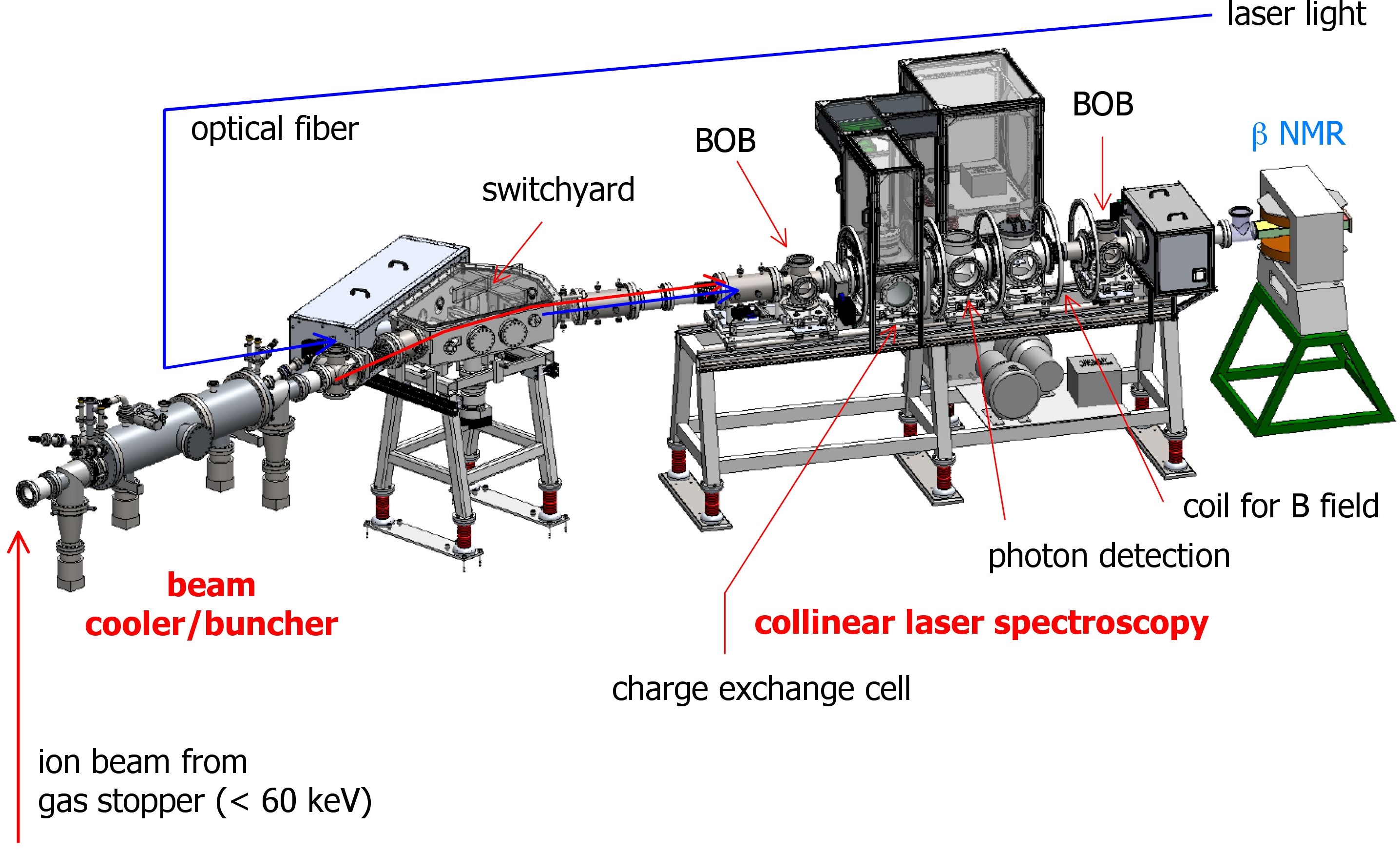
Figure 1: BECOLA beam line 60 keV beam enters from left and proceeds through the beam line.
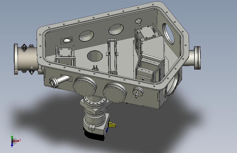
Figure 2: The two-way bending section of the BECOLA beam line. The ion beam enters from the left, is deflected by a total of 30 degrees, and exits to the bottom-right.
Two quadrupole doublets are used to tune the ion beam through the collinear laser spectroscopy beam line. The ion beams may subsequently be passed into a charge exchange cell (CEC), as shown in figure 3, where an alkali vapor (e.g. sodium) will be used to neutralize the ion beam via charge exchange reactions [1,2]. The necessity of neutralization depends on availability of atomic transitions accessible with the laser system. The CEC will be on the variable potential to permit scanning of the ion beam velocity to search for atomic transitions with constant laser frequency [3]. The Doppler shift of laser frequency due to the collinear geometry makes the ion-velocity scan equivalent to laser-frequency scanning.
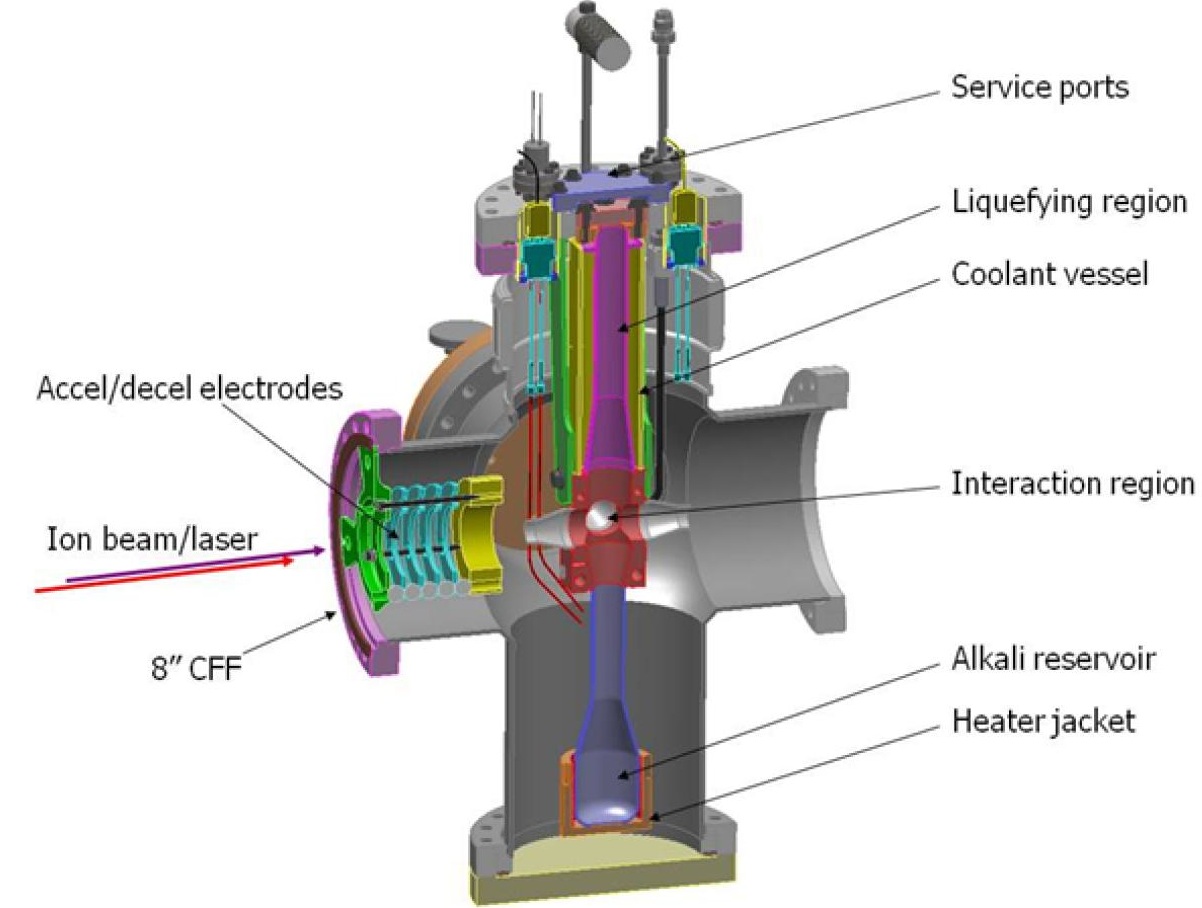
Figure 3: Isometric view of the charge-exchange cell.
The hyperfine structure is determined through measurements taken in the photon detection system (figure 4). Two photomultiplier tubes (PMTs) are used, depending on the wavelength range: one for near-UV light (350 – 500 nm), and one for near-IR light (700 – 1000 nm). The collinearly-propagated laser and ion/atom beam pass through one focal point of the ellipsoidal reflector, and the fluorescence emitted by the beam gathers at the second focal point. In the near-UV setup, said fluorescent photons are immediately captured by the head-on PMT, while in the near-IR setup, the photons are transported from the second focal point to the active area of the side-on PMT via a light guide. The PMT re-expresses the photon count as a readable electrical signal. By measuring the fluorescence of atom/ion beam as a function of the effective laser frequency, the hyperfine spectrum of the atom/ion can be obtained. By knowing the locations of resonances, such nuclear properties as dipole moment, quadrupole moment, and mean square charge radius can be extracted.
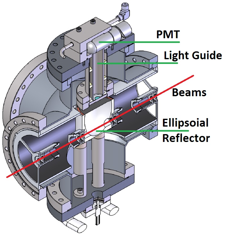
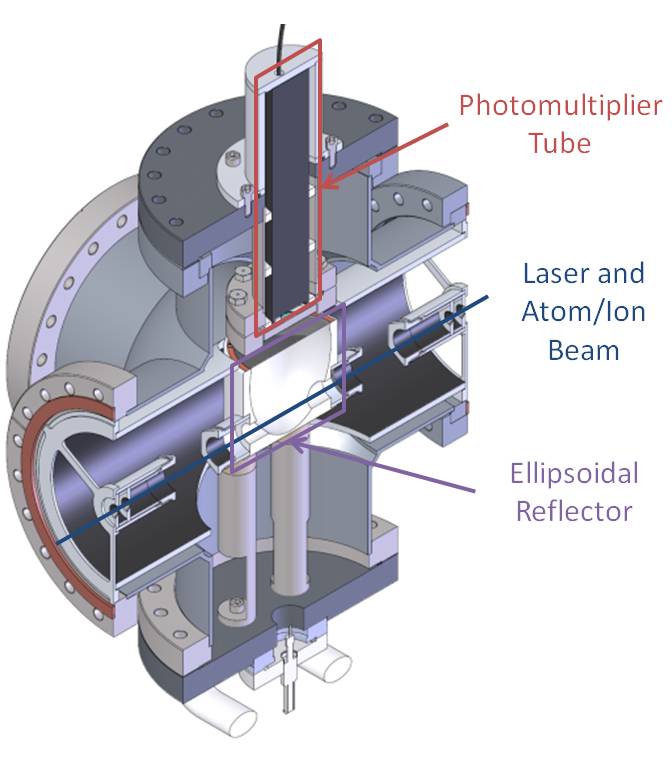
Figure 4: Photon Detection System for near-IR light (above, upper) and near-UV light (above, lower). Note the differing placement of the photomultiplier tube.
Another laser-excitation technique employed for experiments utilizing the BECOLA facility is optical pumping. The ion/atom beam will interact with laser light collinearly over a distance of approximately 1.5 m. Circularly polarized light will be used for optical pumping to produce atomic polarization. A guiding weak magnetic field (~10 G) will be applied along the interaction region to define the quantitation axis for the hyperfine substates. The hyperfine selection rules for circularly polarized photons exciting electrons across hyperfine levels are such that the change in the magnetic substate must be +1 (right-handed) or -1 (left-handed). After many excitations and subsequent relaxations, the beam is atomically polarized. This concept is depicted in figure 5. The polarized beam will then be implanted into a single crystal placed at the center of a dipole magnet for Beta-ray detecting Nuclear Magnetic Resonance (Beta-NMR). Due to external strong magnetic field, the hyperfine interaction between valence electron and nucleus is decoupled and pure nuclear states can be detected. Fundamental properties of the nucleus, such as ground state magnetic dipole and electric quadrupole moments, can be determined.
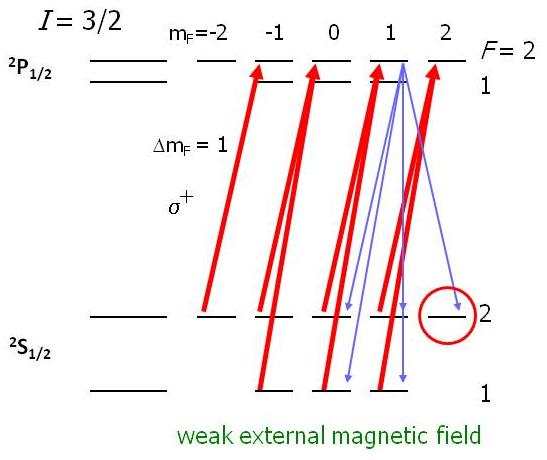
Figure 5: Concept of optical pumping, after many laser-excitations, the m_F = +2 state for the ground state will be preferentially populated.
[1] K.-R. Anton et al., PRL 40,642 (1978).
[2] N. Bendali et al., JPB 19, 233 (1986).
[3] A. Klose, et al., Nucl. Instrum. Methods A, 678, 114 (2012).